Miserable Malalignment Syndrome – is that really what it is called?
Yes, Miserable Malalignment Syndrome (MMS) is a real condition. It is also known as Torsional Malalignment Syndrome (Leonardi et al., 2014). Pain and discomfort are common in those who endure this condition, therefore “miserable” is an apt description.
Miserable Malalignment Syndrome is a diagnosis that may occur in individuals with hip dysplasia or it may occur in individuals with typical hip development. In MMS, the leg bones are rotated in or out more than normal. The femur and tibia are the bones that are typically affected (Sanders Clinic, n.d.). The femur (thigh bone) is the long bone in your leg that connects the hip to the knee. The tibia (shin bone) is the long bone that connects your knee to your ankle. These rotational deformities can be seen in individuals with hip dysplasia and other complex hip diagnoses, but they may also be seen in isolation.
What Causes Miserable Malalignment?
Internal rotation of the leg bones is typical in infants and young children, but the bones usually straighten (“de-rotate”) to adult values through joint loading and with growth (Boston Children’s Hospital, 2023). There is a typical range of rotation in the femur and tibia. In some people, the bones do not straighten or end up rotated too far outward and result in malalignment. It is important to note that normal ranges of rotation in the femur and tibia can vary depending on how the measurement is performed and what measurement tools or technology is used. Diagnosis and treatment recommendations depend on multiple factors including history, symptoms, physical examination, function, and imaging.
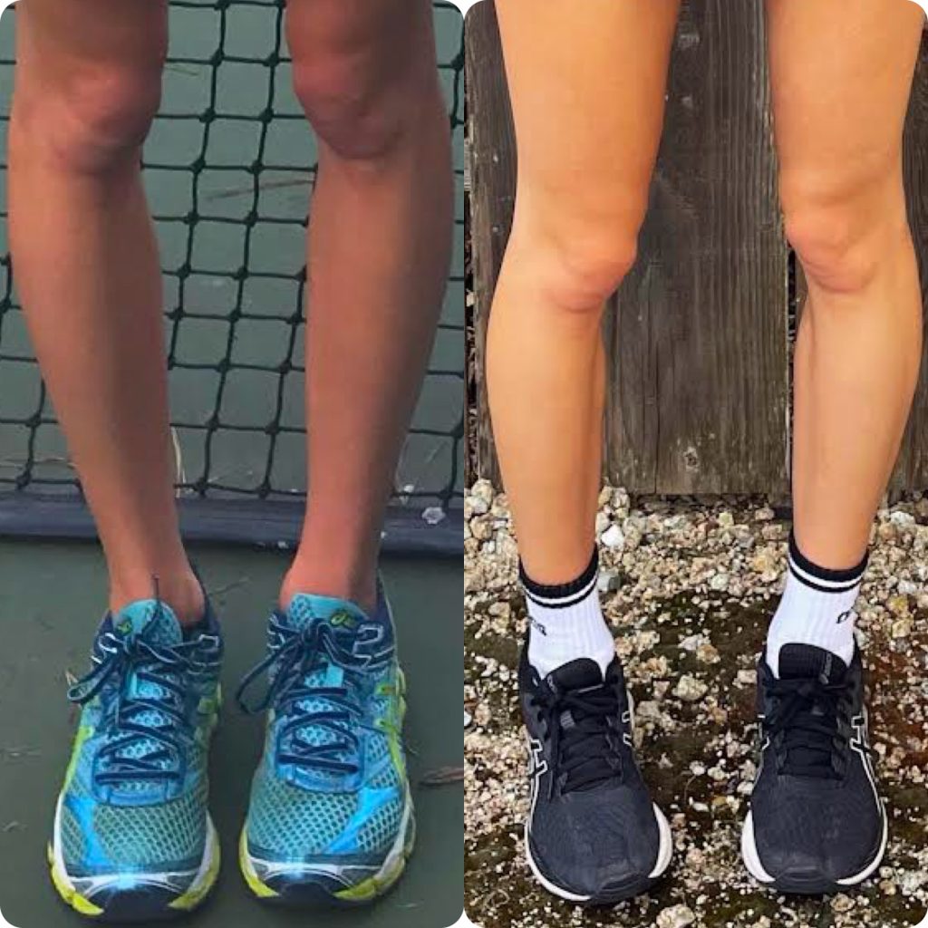
Rotational abnormalities can occur when the femur and/or tibia are rotated too far outwards (external rotation) or inwards (internal rotation). When the femur is externally rotated, it is called femoral retroversion or retrotorsion (Hospital for Special Surgery, 2019). When the femur is internally rotated, it is referred to as femoral anteversion or antetorsion (Hospital for Special Surgery, 2019). Abnormal rotation in the tibia may be described as excessive external tibial torsion or internal tibial torsion. A common presentation for miserable malalignment is when the femurs are rotated too far inward (femoral anteversion) and the tibias are too far outward (external tibial torsion). People with this presentation may have their kneecaps facing inward if they walk with their toes pointing forward or they may walk with their kneecaps straight but with increased out-toeing.
What are some signs or symptoms of MMS?
Miserable malalignment can cause pain, stiffness, and/or clicking in the hips, knees, and ankles (Gruskay, Fragomen, and Rozbruch, 2019, p. 1). A person may experience pain or asymmetries in other parts of the body (such as one shoulder being higher than the other) as it compensates for these conditions. In MMS, the femur and the tibia bones may not move in a coordinated way when a person stands or moves. According to Teitge (2018), “Because the femur and tibia are twisted, the axes of the hip joint, knee joint, and ankle joint do not line up in the same plane.” This causes the femur and the tibia to move in opposite directions. In some cases, it may also cause “maltracking” of the patella (kneecap) and can lead to kneecap subluxations (bone moves partially outside its typical position) or dislocations (bone moves fully outside its typical position). Abnormal rotation in these bones can cause in-toeing or out-toeing (Boston Children’s Hospital, 2023). The in-toeing or out-toeing may be related directly to the rotation in the bone or may be related to compensations for abnormal bone rotations. Walking with in-toeing or out-toeing is not the only indicator of MMS and can be seen with other conditions in the leg as well.
Miserable Malalignment is diagnosed by an orthopedic doctor. The doctor will talk to you about your symptoms and will perform a physical examination to learn about your alignment, joint mobility, and strength. They may look at the way you walk (your gait pattern) and how you move during activities like running, squatting, or balancing on one leg. They may recommend imaging studies like x-rays, MRIs (magnetic resonance imaging), and CT (computerized topography) scans. These will help your doctor learn about your alignment, overall joint health, any injuries to the muscles, tendons, ligaments, or cartilage in or around the joint, and measures of your bone rotations. Since MMS is a complex diagnosis, it may be helpful to find an orthopedist who has experience or expertise working with patients with MMS and similar diagnoses.
How is MMS Treated?
Conserative Management:
Conservative, non-surgical management is often recommended as a first line of treatment and may help reduce pain and improve function. Conservative management may include rest or activity modification and refraining from activities that cause pain, bracing, pain-reducing modalities like ice or heat, and pain medications. Physical therapy to strengthen the leg and core muscles can reduce pain and improve function by taking some of the pressure off of the joints itself (Riley J. Williams III, MD, 2024).
Surgical Management:
People who do not have significant improvement in symptoms and function with conservative management may be candidates for surgical management for MMS. Surgery for MMS is often recommended in older children and adults who are “skeletally mature” or close to being skeletally mature since bony rotations change during growth and may improve without any formal intervention into school-age/early adolescence. If surgery is done before a child is close to being done growing, there may be a risk of over-correcting the rotation or the bone remodeling back to where it was after surgery. Surgery for MMS often includes rotational osteotomies, which are surgeries where bone cuts are made and the bones are then rotated to realign them. The typical osteotomies for Miserable Malalignment Syndrome are femoral osteotomies and tibial osteotomies. A femoral osteotomy is when the femur/thigh bone is cut and rotated, and a tibial osteotomy is when the tibia/shin bone is cut and rotated. When the bones are rotated to address alignment, there will be a gap between the cut in the bone.
Hardware is used to hold the bones in place until the gap fills in and the bone heals. Your surgeon will determine the best hardware to stabilize the bones based on the type of surgery, patient characteristics, and their experience. Hardware can be external (outside the body) or internal (inside the body) (Matthew et. al, 2011). An external fixator is a includes an outside frame that has pins attached that go inside the leg to stabilize the bone (Matthew et. al, 2011). An internal fixator is typically either a plate and screws or a rod and screws that go inside of the leg. Hardware may be removed through a second procedure months or years after the osteotomy when bone healing is complete. Some surgeons regularly remove hardware and others will only remove it if the patient is having pain or other symptoms from it. You should discuss the risks and benefits of hardware removal with your surgeon.
Your surgeon will decide if you need a distal or proximal osteotomy. Distal means that the cut will be made farther away from the center of the body. For example, a distal femoral osteotomy means that the cut is made closer to the knee rather than closer to the hip. Proximal means that the cut will be made closer to the center of the body. For example, a proximal femoral osteotomy means that the cut is made closer to the hip than the knee. Distal versus proximal is decided upon a few factors, such as the surgeon’s preference and where the pain is located (Niklasch et. al, 2018).
What is Recovery Like After Surgery for MMS?
Many patients are not allowed to put full weight through their leg after surgery for MMS or will be limited due to pain and discomfort after surgery. A wheelchair, walker or crutches may be required for several weeks or months after surgery until you are allowed to put full weight through your leg and can safely and comfortably walk without significant pain or limping (Sheth, Manner, Foran, 2022). Depending on the location of the surgery and the surgeon’s preference, you may need to use a brace or a cast for protection while the bone heals (Sheth, Manner, Foran, 2022). Physical therapy will be helpful after surgery regain joint motion, flexibility, and muscle strength, to retrain walking, and return to daily activities, work, school, and recreational activities (Sheth, Manner, Foran, 2022). Surgeons may have different recovery guidelines and protocols, and these will also be based on individual patient characteristics and healing. Weight-bearing, precautions, bracing and casting, and timing of physical therapy and activity progression can all vary from patient to patient and among surgeons, so be sure to check with your surgeon to get the specifics for your surgery and recovery. It is important for patients to partner with their surgeon and physical therapist, follow post-operative guidelines, and give themselves time and patience to progress safely and properly to have a timely and positive recovery.
Conclusion
Miserable Malalignment Syndrome can be a complex and painful condition. Although MMS is not a widely known diagnosis, there are resources and health professionals that can guide you in this process. An informed and skilled care team is integral in making a diagnosis, the rehabilitative process, and even mental health support, as chronic pain can be stressful on the body and mind. Additionally, reaching out to others who have MMS to learn about their experience may be helpful. There is a MMS patient support group on Facebook and others have shared their journeys through individualized social media accounts. You can also ask your surgeon or other healthcare providers if they have any patients who they have treated who would be willing to serve as patient peer mentors. However, it is important to remember that every patient is unique and your experiences, treatment plan, and outcomes may be quite different from others you meet. Make sure to discuss all questions and concerns about your unique case with your surgeon and healthcare team so you can work together to figure out the best treatment for you.
Author: Kait Williams
Citations:
Boston Children’s Hospital. (2023). Tibial torsion. Tibial Torsion | Boston Children’s Hospital. https://www.childrenshospital.org/conditions/tibial-torsion
Gruskay J.A, Fragomen A.T, Rozbruch S.R. (2019). Idiopathic Rotational Abnormalities of the Lower Extremities in Children and Adults. JBJS Rev. 2019 Jan;7(1):e3. doi: 10.2106/JBJS.RVW.18.00016. PMID: 30624306.
Hospital for Special Surgery. (2019, September 19). Hip/femoral anteversion: Causes, symptoms, treatment. https://www.hss.edu/condition-list_hip-femoral-anteversion.asp
Leonardi F, Rivera F, Zorzan A, Ali S.M. Bilateral double osteotomy in severe torsional malalignment syndrome: 16 years follow-up. J Orthop Traumatol. 2014 Jun;15(2):131-6. doi: 10.1007/s10195-013-0260-0. Epub 2013 Aug 29. PMID: 23989854; PMCID: PMC4033816.
Matthew Seah, K. T. BMedSci, MBChB; Shafi, Raheel MD; Fragomen, Austin T. MD; Rozbruch, Robert S. MD. Distal Femoral Osteotomy: Is Internal Fixation Better than External?. Clinical Orthopaedics and Related Research 469(7):p 2003-2011, July 2011. | DOI: 10.1007/s11999-010-1755-0
Niklasch, M., Boyer, E.R., Novacheck, T., Dhreher, T., Schwartz, M. (2018). Proximal versus distal femoral derotation osteotomy in bilateral cerebral palsy. Developmental Medicine and Child Neurology. 60(10), 1033-1037. https://doi.org/10.1111/dmcn.13910
Riley J. Williams III, MD, Manhattan Hip, Knee, and Shoulder Specialist| Hospital for Special Surgery (2024). Knee Malalignment Syndrome. https://rileywilliamsmd.com/knee-miserable-malalignment-syndrome-manhattan-new-york-city-ny/
Sanders Clinic (n.d.). Rotational deformity malalignment disorder. https://sandersclinic.net/rotational-deformity-malalignment-disorder/
Sheth, N.P., Manner, P.A., Foran, J.R.H. (2022). Osteotomy of the knee – orthoinfo – aaos. OrthoInfo. American Academy of Orthopaedic Surgeons https://orthoinfo.aaos.org/en/treatment/osteotomy-of-the-knee/
Teitge RA. (2018). The power of transverse plane limb mal-alignment in the genesis of anterior knee pain-clinical relevance. Ann Joint 2018;3:70.