Hip dysplasia experts may order a variety of special imaging and tests to help determine the best treatment for a patient. These are some of the most common measurements associated with evaluation of hip dysplasia. It is important to keep in mind that measurements are not always black and white, and may vary slightly between different providers (“inter-rater reliability”) and sometimes even within the same provider on multiple occasions (“intra-rater reliability”). A good hip dysplasia expert will not make a treatment plan based solely on measurements. Instead, he or she will combine information from the patient’s history, physical examination, and imaging to determine the best plan of care.
Lateral Center Edge Angle (LCEA)
This is the most commonly measured angle to determine if a person has hip dysplasia. It measures how well the acetabulum (“hip socket”) covers the head of the femur (“ball” of the hip joint).
Normal values are between 25-35 degrees
< 20-25 degrees = hip dysplasia (shallow hip socket)
> 39 degrees = over-coverage of the acetabulum and is associated with pincer impingements in femoral acetabular impingement (FAI)
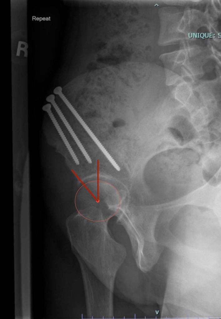
Anterior Center Edge Angle (ACEA)
The ACEA measures how well the front of the acetabulum (“hip socket”) covers the head of the femur (“ball” of the hip joint) on false profile x-rays.
Normal values are between 20-45 degrees
< 20 degrees = hip dysplasia (shallow hip socket)
> 45 degrees = over-coverage of the acetabulum and is associated with pincer impingements in femoral acetabular impingement (FAI)
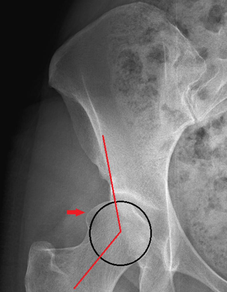
Alpha Angle
The head of the femur should round. The alpha angle measures how much the shape of the femoral head differs from the normal spherical shape.
> 55 degrees is associated with CAM morphology that can be seen in femoroacetabular impingement and hip dysplasia. Notice how there is extra bone outside the circle (red arrow).
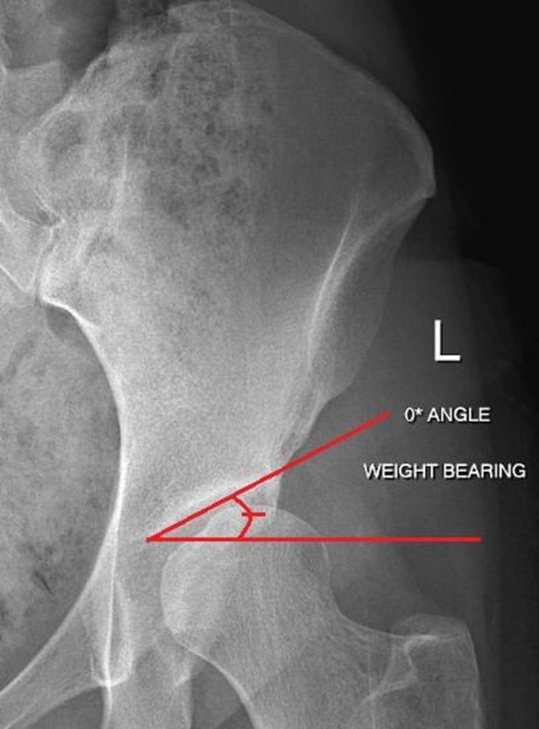
Tonnis Angle
The Tonnis angle measures the upward slope of the hip socket. A slope between 0-10 degrees is normal and allows for even loading across the joint.
>10 degrees: a steep slope may be seen with dysplasia. This increases load/stress placed across smaller parts of the joint, and can cause pain and damage to the cartilage.
< 0 degrees: a shallow slope may be seen with FAI. This can cause pain and may limit hip range of motion.
– A slope between 0-10 degrees is normal and allows for even loading across the joint.
Acetabular Index / Angle of Sharp
Hips with dysplasia have increased forces along the weight-bearing parts of the hip socket. This can cause cartilage damage and early arthritis. An increased acetabular index is seen with dysplasia and results in increased forces being loaded across a smaller surface area which causes increased pressure and stress in the joint.
> 42-45 degrees = acetabular dysplasia
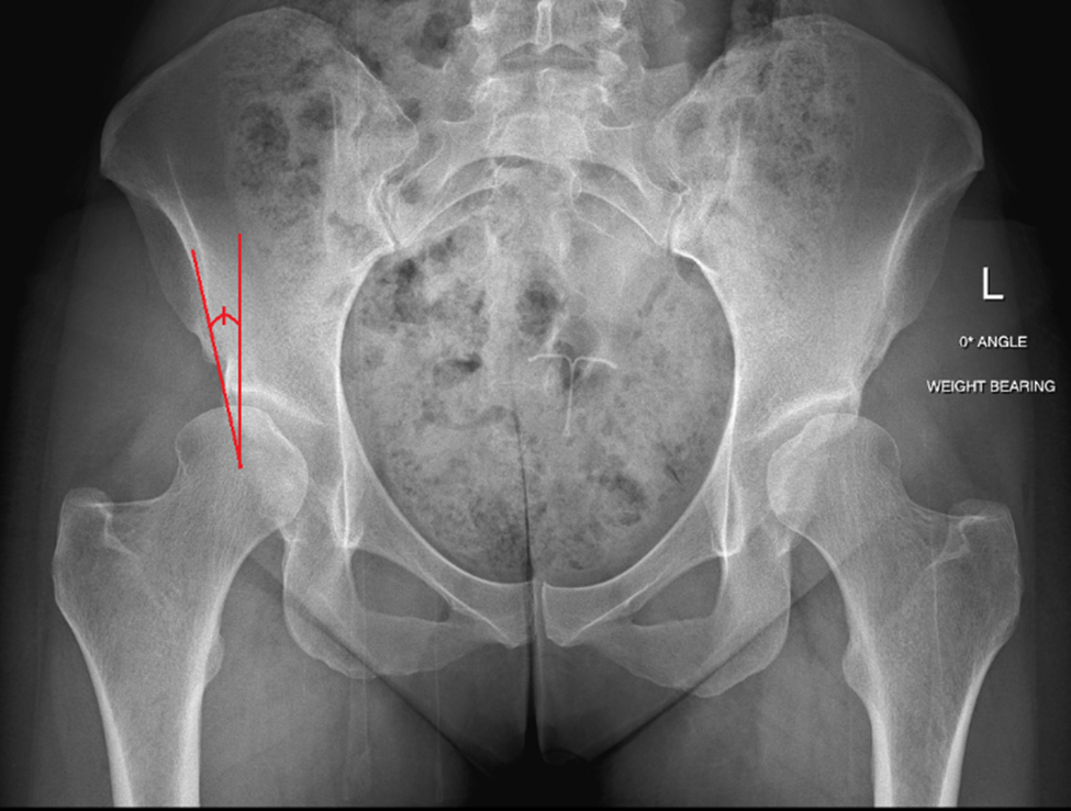
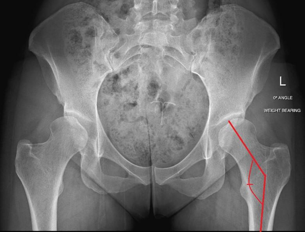
Femoral Neck-Shaft Angle
This angle is formed by the line going from the center of the head of the femur down the middle of the “neck of the femur” (this is the part of the bone that connects the head (ball) of the femur to the long bone of the femur (shaft)). This measurement is used to diagnose hip conditions like hip dysplasia, FAI, Legg-Calve-Perthes disease, avascular necrosis, osteogenesis imperfecta, and fractures up near the top of the femur. This angle may be too high or too low in people with hip problems.
Normal angle =125-135 degrees
< 125 degrees = “coxa vara” (“varus”)
> 135 degrees = “coxa valga” (“valgus”)
References:
https://www.ncbi.nlm.nih.gov/pmc/articles/PMC5193516
https://www.sciencedirect.com/science/article/pii/S1063458413010121
https://www.sciencedirect.com/science/article/pii/S2212628717301767
https://www.hindawi.com/journals/bmri/2016/8645027
Hanson JA, Kapron AL, Swenson KM, Maak TG, Peters CL, Aoki SK. Discrepancies in measuring acetabular coverage: revisiting the anterior and lateral center edge angles. J Hip Preserv Surg. 2015 Jun 13;2(3):280-6. doi: 10.1093/jhps/hnv041. PMID: 27011850; PMCID: PMC4765297.
Cheng H, Zhang L, Luo D, Ren N, Zhang Z, Gu W, Hu Y, Zhang H. Determining anterior hip coverage in patients with hip dysplasia using the anterior center-edge angle on Lequesne’s false-profile radiograph and on computed tomography. J Hip Preserv Surg. 2023 Mar 6;10(1):42-47. doi: 10.1093/jhps/hnac048. PMID: 37275833; PMCID: PMC10234381.
Author: Ashley Imgrund-Flora, MS, PA-C
Updated March 2026, Edited & Reviewed by Lauren Schoeller, MS & Wudbhav N. Sankar, MD
Disclaimer: The information on the Miles4Hips website is meant for informational purposes only. While our goals are to promote understanding and knowledge of hip dysplasia and to empower patients and their families in healthcare decision making, we cannot guarantee accuracy or appropriateness of the information for your specific condition or circumstances. The information on this site is not meant to take the place of the professional judgment of your medical providers. Individuals should always seek the advice of your physician/surgeon, physical therapists, and other qualified health care provider with any questions you may have regarding a medical condition or treatment. Individuals should never disregard the advice of your medical providers or delay in seeking it because of something you have read on this website.